Description
FEATURES
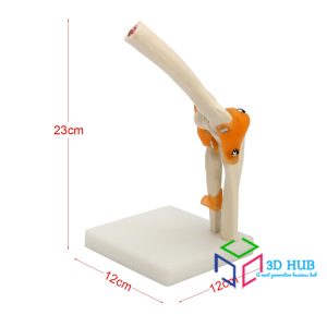
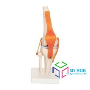
Life-Size Human Knee Joint Anatomy Model
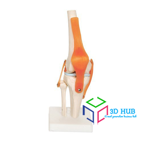
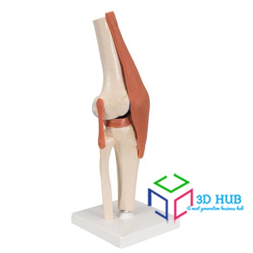
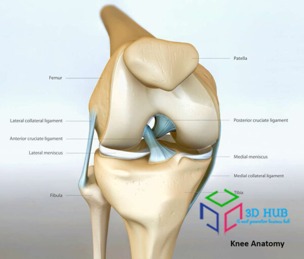
The Life-Size Knee Joint Model features 18 bones, ligaments, and tendons involved in the knee joint’s anatomy. Cast from real human bones, the model includes the fibula, tibia, patella, femur and the associated bony landmarks and textures.
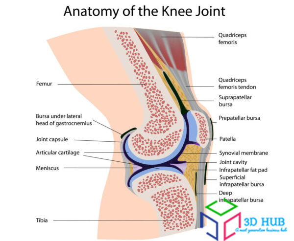
Our knee joint model offers an anatomically correct representation of the connection between the different bones in the area and how they relate to the anatomy, structure, and stability of the knee. As one of the strongest and most complex joints in the human body, the knee joint can support the body’s weight when upright and help propel the body forward when walking. This anatomy model features the patella (kneecap) which provides articulation to the femur.
Knee Ligaments
This knee joint anatomy model features textured ligaments to provide a better understanding of the connective structures around the bones. The tough elastic tissues are presented prominently on the model to represent a detailed view of the human knee joint’s anatomy.
The human knee joint is susceptible to several injuries. These injuries can be from common, daily events or athletic in nature. The ligaments represented on the model includes the anterior cruciate ligament (ACL) and the posterior cruciate ligament (PCL), both major ligaments that provide stability to the knee joint. Tears and sprain commonly occur in these ligaments, most notably in sports. This knee joint with ligaments model allows for the study of how these ligaments co-exist with the other ligaments, tendons, and bones in the knee.
Sturdy Base Stand
Our knee joint with ligaments model comes with a sturdy base stand, making it perfect for study, display, and transport. The base stand allows the model to be viewed upright for an uninterrupted study of the knee joint anatomy.
Full-Color Detailed Study Guide Booklet & User Guide
The Life-Size Human Knee Joint with Ligaments Model comes with a detailed full-color study guide booklet that identifies the 18 bones, ligaments, tendons, and meniscus bands presented on the skull model.
PRACTICAL APPLICATIONS: Perfect for students, medical practitioners, professionals, and artists.
Students
Medical students can get premium knowledge of the anatomy of the human knee joint and its ligaments. This knee joint model is life-size and features realistic textures, bony landmarks, and a desktop stand, making it a perfectly compact classroom or home study tool. This anatomy model and the accompanying study guide are helpful tools to prepare for medical school education and future medical practice.
Orthopedists and Orthopedic Surgeons
Medical professionals in the orthopedic department can find this life-size knee joint with ligaments model useful for patient demonstration in order to expand their understanding of knee function, injuries, surgeries, and medical processes.
Physical and Occupational Therapists
The model provides an anatomically accurate depiction of the knee joint and its ligaments. This knee model is useful for patient demonstration of correct joint positioning, movement, and stability.
Sports Medicine and Athletes
Professionals in sports and sports medicine can gain the correct visualization and demonstration of positioning and movement in the knee joint. The anatomically correct knee joint model is a helpful tool in reviewing the human shoulder for monitoring and diagnosing common athletic injuries like torn ACLs and PCLs.
Artists and Anatomists
Produce high-quality anatomical art with our knee joint and ligaments model. The realistic details, textures, landmarks, and flexibility are great tools for understanding the basics of human anatomy for artistic endeavors.


Reviews
There are no reviews yet.